
- M/63
- 경비직
- hypertension, diabetes, hyperlipidemia
- old CVA Hx(+): cerebral infarction (2022. 5.)
- last normal time: 3일 전
- 친동생에게 전화가 왔으나 말을 하지 않았다. 급하게 가 보니 말을 제대로 하지 못하고, 제대로 움직이지도 못해 응급실로 내원
- confusional mentality
- dysasthria, Lt. eyeball deviation, obey 일부 가능
- motor G4
- Lab) Hb 12.1, Hct 35.5%, WBC 11000, CRP 6.9, AST/ALT 71/42, CPK 963.7, BUN/Cr 39.1/1.1, serum glucose 155, PT/aPTT 10.5/17.6, INR 0.95, Na-K-Cl 134.8-3.94-101.5, CK-MB/TnI 4.3/0.01, ProBNP 1310
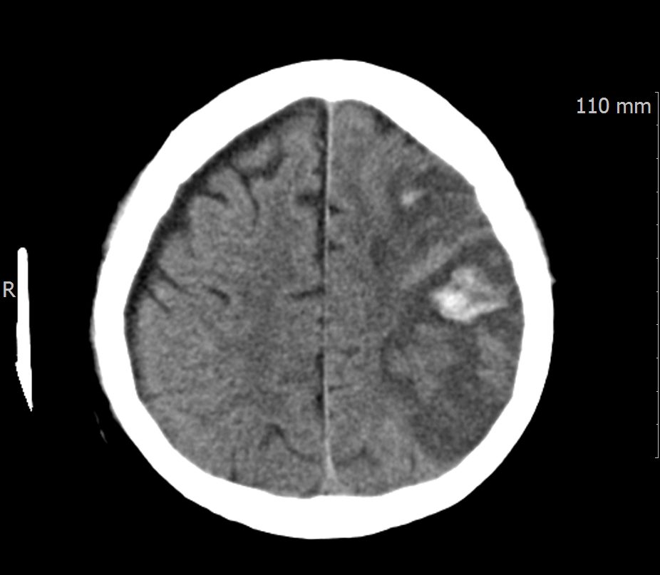
- Brain CT)
- hemorrhagic conversion of left MCA territory subacute infarct.
- r/o) chronic infarct in right frontal cortex with encephalomalacia.
- r/o) chronic SDH in right fronto temporo parieal lobe , ddx) subdural hygroma.
- post op fixation of left maxilla, left zygoma.
- r/o) old fx of medial wall of both orbit.


- 진단
- #1. subacute cerebral infarction
- #2. hemorrhagic conversion
Hemorrhagic transformation of ischemic stroke
- 동맥 폐쇄로 인한 급성 뇌경색에서 출혈 변환이 18~42% 정도 발생한다고 한다.
- 현미경 검사에서 창백경색(pale infarct)으로 분류된 환자의 상당수도 출혈점(petechiae)이 존재한다. 출혈의 정도와 범위가 다를 뿐 대개 뇌경색 환자의 출혈은 존재한다고 볼 수 있다.
- 이러한 출혈변환의 기전은 허혈과 재관류 시에 진행되는 BBB의 파열이다.
- 출혈변환은 크게 점출혈이 보이는 출혈경색(hemorrhagic infarct)과 종괴효과(mass effect)를 동반하는 실질혈종(parenchymal hematoma)로 나눌 수 있다.
- 단순히 출혈변환이 존재하는 것이 중요한 것이 아니라, 증상의 악화를 유발하는가가 중요하다. 즉, 유증상 출혈변환과 무증상 출혈변환으로 생각하는 것이 좋다.
🔸ECASS II Criteira
- Hemorrhagic Infarction type 1 (HI1): petechial hemorrhages at the infarct margins
- Hemorrhagic Infarction type 2 (HI2)
- petechial hemorrhages throughout the infarct
- no mass-effect attributable to the hemorrhages
- Parenchymal hematoma type 1 (PH1)
- =< 30% of the infarted area
- minor mass effect attrubutable the the hematoma
- Parenchymal hematoma type 2 (PH2)
- > 30% of infarct zone
- substantial mass effect attributable to the hematoma
🔸출혈변환의 메카니즘

Possible mechanisms in early and delayed HT. The disruption of the BBB is a common pathway in HT formation following acute ischemic stroke. Various molecules from neutrophils and peripheral blood in possible processes in early HT are mainly associated with HT after ischemic stroke. Exogenous tPA can also increase MMP-9 levels by activating neutrophils and increasing MMP-2 levels. Conversely, in possible processes for delayed HT, the brain tissue is a major source of MMP-9 within the first 18–24 h following stroke, and endogenous tPA can act on endothelial cells to increase MMP-2 release from astrocytes as well as MMP-9 release from microglia. HT, hemorrhagic transformation; NVU, neurovascular unit; MMP, matrix-metalloproteinase; ROS, reactive oxygen species; tPA, tissue plasminogen activator; BBB, blood-brain barrier; VEGF, vascular endothelial growth factor.
(출처: NIH – https://pmc.ncbi.nlm.nih.gov/articles/PMC8669478/figure/F2/)
- MMP-9는 BBB의 파괴, 비정상적인 투과성을 증가시키는 원인
- 외인성, 내인성 tPA는 MMP-2를 증가
- MMP-2는 미세아교세포(microglia)에서 MMP-9 방출을 증가
- MMP-9는 기저층 IV형 콜라겐의 파괴에 중요한 역할을 하여 혈관성 부종을 유발하고 HT의 위험성을 높힌다.
🔸뇌졸중의 출혈변환 위험을 증가시키는 요인
- 노인
- 큰 경색 범위
- 심장성 혈전 뇌졸중
- 항응고제의 사용
- 발열
- 고혈당
- 낮은 콜레스테롤 수치
- 수축기 혈압의 상승
- 혈전용해제 치료 또는 다른 재관류 치료
🔸정맥 혈전용해(intravenous thrombolysis) 후 출혈성 변화의 예측인자
- 중증의 경색 (NIHSS > 14)
- proximal MCA 폐쇄
- MCA territory의 1/3 이상 범위에서 CT의 hypotensity lesions
- 경색 발생 후 6시간 이상 지연된 재관류
- 빈약한 곁순환(collateral flow)
🔸치료

Treatment algorithm for appropriate medical and surgical approaches to HT after ischemic stroke. NIHSS, National Institutes of Health Stroke Scale; CT, computed tomography; MRI, magnetic resonance imaging; ICU, intensive care unit; LMWH, low molecular weight heparin; UFH, unfractionated heparin; SBP, systolic blood pressure; IV, intravenous; PCC, prothrombin complex concentrates; FFP, fresh frozen plasma; INR, international normalized ratio; DOAC, direct oral anticoagulants; EVD, external ventricular drainage; ICP, intracranial pressure.
(출처: NIH – https://pmc.ncbi.nlm.nih.gov/articles/PMC8669478/figure/F4/)
알아두기 위해 발췌해 둔 위 내용들은 NIH에서 가져왔지만, 우리나라 아주대학교병원 신경과에서 연구하여 2021년 11월에 발표한 논문이다.

